Revolutionising Medical Imaging with Generative Adversarial Networks Applications and Innovations
- My Academic Tutor

- Mar 13
- 3 min read
Medical imaging plays a crucial role in diagnosing and treating diseases. Yet, challenges such as image quality, radiation exposure, and data scarcity often limit its effectiveness. Generative Adversarial Networks (GANs) offer promising solutions by enhancing image quality, generating realistic synthetic data, and adapting imaging modalities. This post explores how GANs transform MRI, CT, and digital pathology, highlighting key applications and innovations.

Understanding GAN Architecture

GANs consist of two neural networks: a generator and a discriminator. The generator creates synthetic images, while the discriminator evaluates their authenticity against real images. These networks train together in a competitive process, improving the generator’s ability to produce realistic images over time.
Generator: Produces new images from random noise or latent vectors.
Discriminator: Distinguishes between real and generated images.
Training: Both networks improve through adversarial feedback, leading to high-quality image synthesis.
This architecture allows GANs to learn complex data distributions, making them ideal for medical imaging tasks where data can be limited or noisy.
Latent Space Representation
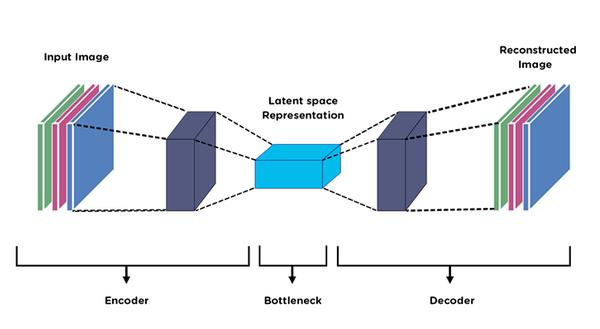
Latent space is a compressed, abstract representation of data learned by the generator. Each point in this space corresponds to a unique image. By manipulating latent vectors, researchers can generate variations of medical images, such as different tissue textures or anatomical structures.
Enables controlled image synthesis.
Supports interpolation between image features.
Facilitates understanding of underlying image characteristics.
In medical imaging, latent space helps create diverse datasets for training diagnostic models or simulating rare conditions.
GAN-Based Medical Image Generation

Generating synthetic medical images addresses data scarcity and privacy concerns. GANs can produce realistic MRI, CT, and histopathology images that augment existing datasets.
Synthetic images improve machine learning model training.
Reduce the need for large, annotated datasets.
Preserve patient privacy by avoiding real patient data reuse.
For example, GAN-generated brain MRI scans have been used to train tumour detection algorithms, improving accuracy without exposing sensitive data.
MRI Super-Resolution Using GANs

MRI scans often face resolution limits due to hardware constraints and scan time. GANs enhance MRI images by increasing spatial resolution, revealing finer anatomical details.
GANs learn to map low-resolution images to high-resolution counterparts.
Improved resolution aids in detecting small lesions or subtle abnormalities.
Faster scans become possible by acquiring lower-resolution images and enhancing them afterwards.
Studies show GAN-based super-resolution can double the effective resolution of MRI scans, supporting better diagnosis and treatment planning.
Low-Dose CT Enhancement

CT imaging exposes patients to ionising radiation, raising safety concerns. Reducing radiation dose lowers image quality, making diagnosis harder. GANs help enhance low-dose CT images by removing noise and artefacts.
GANs restore image clarity while preserving diagnostic features.
Enable safer imaging protocols with reduced radiation exposure.
Support early disease detection through clearer images.
Clinical trials demonstrate that GAN-enhanced low-dose CT images match the quality of standard-dose scans, improving patient safety without sacrificing accuracy.
Histopathology Image Generation

Digital pathology relies on high-quality tissue images for disease diagnosis. GANs generate synthetic histopathology images that mimic real tissue samples.
Augment datasets for training cancer detection algorithms.
Simulate rare pathological conditions for research.
Assist in standardising image quality across labs.
For instance, GANs have produced synthetic breast cancer tissue images that help train classifiers to distinguish malignant from benign samples with higher precision.
Domain Adaptation in Medical Imaging

Medical images vary across devices, protocols, and institutions, causing challenges for AI models trained on one dataset to generalise to others. GANs enable domain adaptation by translating images from one domain to another.
Convert images from different scanners to a common style.
Reduce variability caused by acquisition differences.
Improve AI model robustness across diverse clinical settings.
An example includes adapting MRI scans from different hospitals so a single tumour segmentation model performs well on all data sources.

Comments